
For years, studying cancer cells meant breaking tissue samples apart. Standard lab techniques – Bulk RNA sequencing, proteomics- all require grinding up tissue to analyse individual cells or molecules. It was problematic because you lose all of the cellular context of the tissue, where the cells are, how they’re communicating and what their personal lives were. For decades, grinding tissues were the only option. Now there’s a better way.
What is spatial omics?
Spatial omics is a technology advancement that give researchers the ability to analyse tissue samples (e.g biopsies or surgical resections) without needing to dissociate it. Instead of destroying the tissue structure, these tools map out which cells are where and what they’re doing, all while preserving the biological architecture. It’s the difference between studying individual puzzle pieces versus seeing the whole picture assembled.
How It Actually Works
Several technologies have emerged over the past few years. At Wesley Research Institute’s Queensland Spatial Biology Centre, the team has championed spatial proteomics profiling. This technology can detect dozens of different protein markers on a single tissue slice by staining and imaging in repeated cycles – like taking multiple photographs of the same scene with different filters that image different proteins, each telling us a more deeper biological finding than the last.
What makes this powerful is the type of information you get. Traditional methods give you averages across whole tissue samples or data from isolated cells. Spatial biology gives you a map – showing exactly where each cell type is located, what proteins they’re expressing, and who their neighbours are.
This lets researchers ask questions that weren’t possible before: Are immune cells actually reaching the tumour, or are they stuck at the edges? Are the support cells that feed tumours clustering in areas with low oxygen? Where exactly are the cancer cells that express certain drug targets?
The QSBC’s Ovarian Cancer Discovery
The centre’s work on ovarian cancer shows why location matters. The team mapped out where different immune cells were positioned across tissue samples and found something important: long-term patient survival correlated with how close certain immune cells were to the tumour cells themselves.
It’s not enough to have immune cells present in the tissue, they need to be in the right place to do their job. Think of it like having firefighters in the city versus having them at the building that’s actually burning. The QSBC’s findings suggest that the physical proximity of immune cells, particularly Regulatory T cells and CD8 T Cells, to cancer cells affects how well patients respond to treatment. This is still early-stage research that needs more validation, but it points toward a future where doctors could look at the spatial organisation of a patient’s tumour and predict treatment outcomes more accurately.
Learn more at – https://www.wesleyresearch.org.au/fighter-cells-near-tumours-could-boost-survival-for-ovarian-cancer-patients/
Head and Neck Cancer Progress
A pioneering study from QSBC, recently published in NPJ Precision Oncology, made headlines as one of the largest discovery protein studies in translational cancer research to date. Led by Associate Professor Arutha Kulasinghe, the research marked a world-first in analysing hundreds of proteins and thousands of gene signatures from a single tissue sample.
The study examined samples from 84 patients diagnosed with head and neck squamous cell carcinoma – the seventh most common cancer globally, with nearly 890,000 new cases each year. By studying the tissue architecture in the tumour and adjacent regions, the team found clues that predict if a patient’s treatment will be effective to chemotherapy, or if the head and neck cancer would not respond to therapy.
Learn more at – https://www.wesleyresearch.org.au/world-first-cancer-breakthrough-tests-hundreds-of-clues-in-one-biopsy-to-guide-treatment/
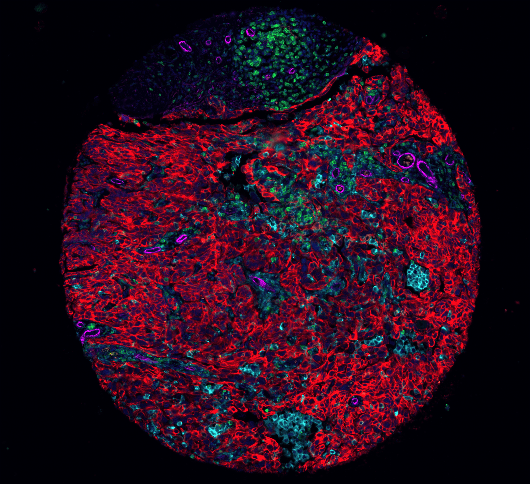
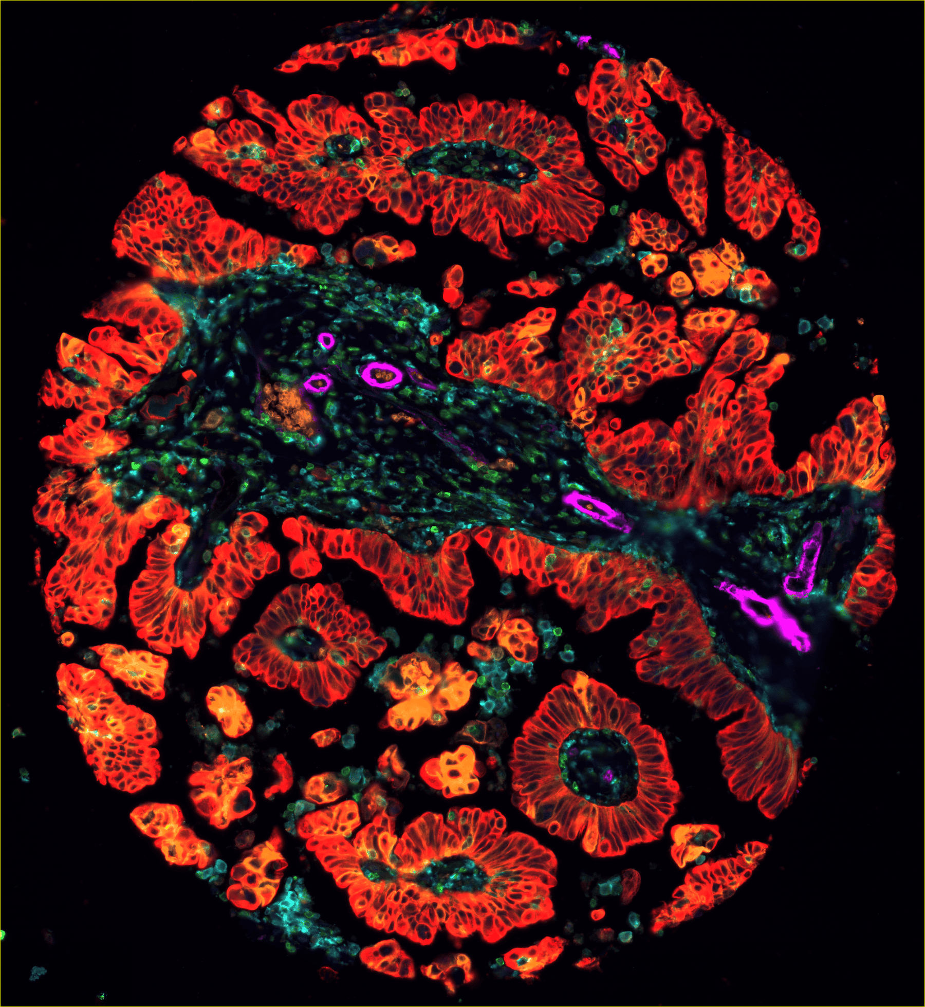
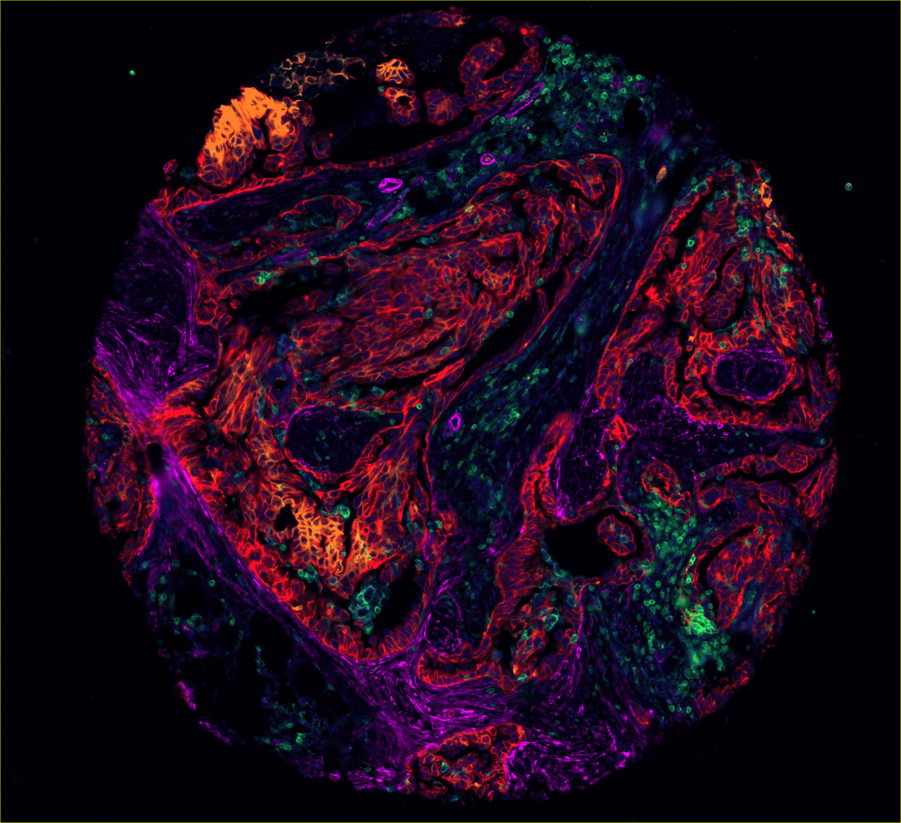
Tissue microarray of 1mm cores from ovarian cancer samples. Red show shows the tumour cells, green and blue show immune cells , purple shows smooth muscle, orange shows areas of the tumour that are metabolically active (taking up glucose).
Personalised Cancer Treatment in Practice
Cancer isn’t uniform. Different regions of the same tumour can behave completely differently. Some areas might be packed with the right types of immune cells ready to fight, some might have exhausted immune cells. Other areas might be “immune deserts” where the body’s defences can’t penetrate. Some regions might have dense supporting tissue, while others are mostly cancer cells.
Immunotherapy – treatments that work by unleashing the immune system against cancer – only succeeds in some patients. Spatial profiling is helping explain why. Associate Professor Arutha Kulasinghe‘s research at the QSBC is mapping these patterns in lung cancer patients. The goal is to identify which patients are likely to benefit from immunotherapy based on how their immune cells are organised around the tumour.
By understanding each patient’s cancer in intricate detail, where we have x,y and z coordinates for cells in space, we can build tissue maps across disease cohorts to identify cellular patterns associated with clinical outcomes. These biomarkers or tissue signatures can be valuable to pathologists and medical oncologists to determine which therapies are most likely to work for individual patients.
Associate Professor Arutha Kulasinghe
What Happens Next
Spatial omics will gradually imbed itself into cancer care over the next 5-10 years, especially as technology and computational learnings evolve. Certain cancer types where spatial information proves most valuable will adopt it first. Widespread use depends on completing validation studies, getting regulatory approval for spatial biomarkers, and convincing health systems that the added cost delivers better patient outcomes.
The technology itself will keep improving – higher resolution, more markers measured at once, even 3D mapping of tumours. But technology isn’t the bottleneck. The computational analysis and making sense of the puzzle are.
Spatial omics isn’t replacing current diagnostic tests. It’s adding a missing piece – the map that shows where everything is located and how cells interact in their actual tissue environment . Combined with genetic and molecular testing, this gives a fuller understanding of each patient’s cancer. How that leads to improved treatment outcomes is what ongoing research is currently determining. The early results are promising, but there’s still substantial work ahead before spatial profiling becomes a standard part of personalised cancer treatment.
Learn more about the Queensland Spatial Biology Centre.