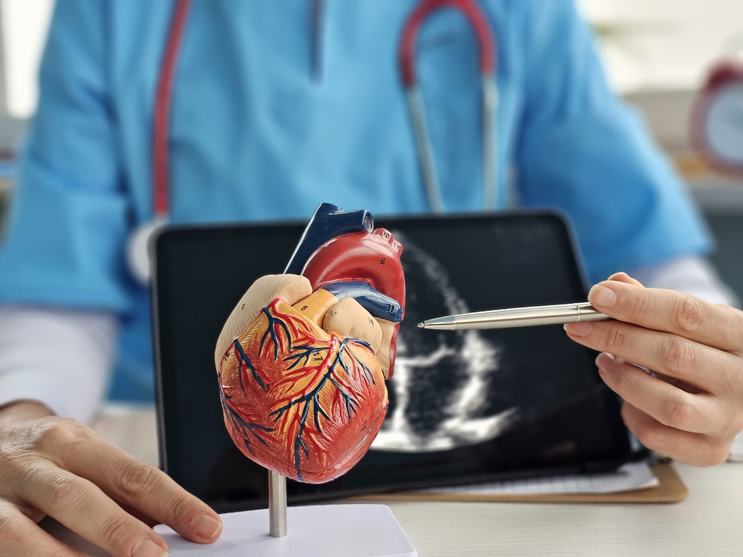
Cardiovascular Research Program
A new research program bringing unique human tissue models, advanced spatial biology analyses, and ‘AI’ tools into cardiovascular research.
Cardiovascular diseases remain the leading cause of mortality worldwide.
Despite decades of progress, cardiovascular care still struggles to account for the enormous variability between patients. Two individuals with the same diagnosis may respond very differently to identical treatments, yet current clinical pathways rarely reflect this complexity.
Dr Eliot Peyster, Head of Cardiovascular Research at the Queensland Spatial Biology Centre (QSBC) is working with our team of exceptional researchers to help bridge this gap by rethinking cardiovascular care. Using unique human tissue models, advanced spatial biology analyses, and ‘AI’ tools, this research program is working to identify biological signatures that predict risk, treatment response, and disease progression, with the goal of moving beyond one-size-fits-all treatment pathways.
About Dr Eliot Peyster
Saving More Lives Through Better Donor Heart Selection
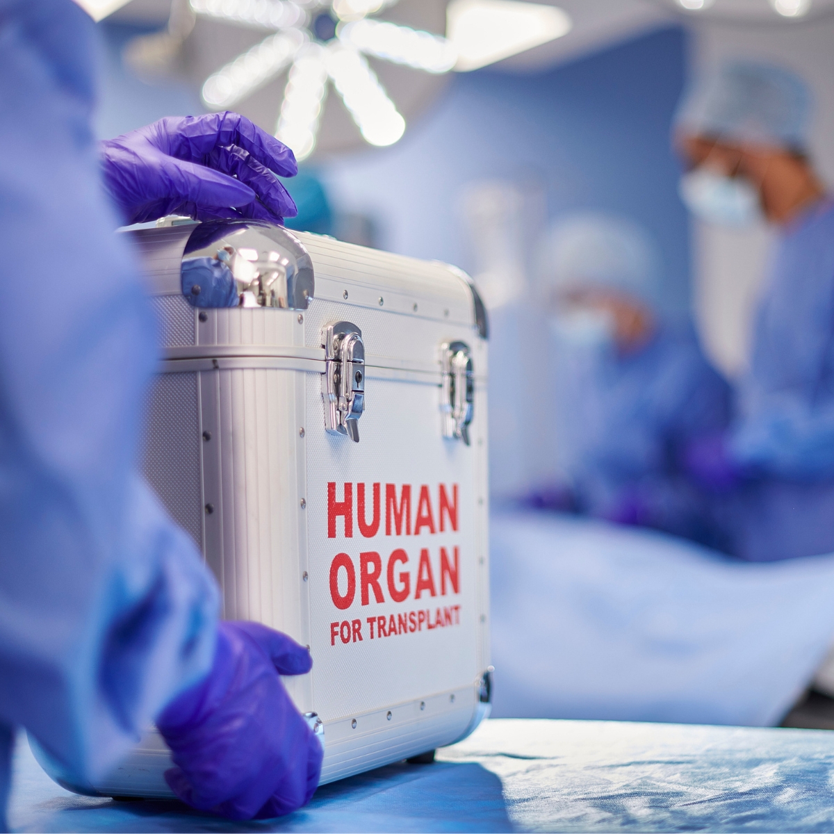
Our research is focused on changing that. By combining advanced imaging of heart tissue with detailed molecular analysis, we are developing a new way to assess donor heart quality – one that goes far beyond the traditional “yes or no” judgement. Early results suggest that many hearts previously considered too risky may in fact be suitable for transplant. With further development, this approach could help doctors make more confident decisions, increase the number of usable donor hearts, and ultimately save more lives.
Finding New Targets in the Silent Phase of Coronary Heart Disease
Our team is taking a different approach. Using human heart tissue from donors with varying degrees of coronary disease, we are studying how the heart changes before severe dysfunction appears. We have found evidence that even earlier-stage coronary disease is associated with important changes in the heart’s small blood vessels, supporting tissue, and energy-producing machinery. These findings suggest that coronary artery disease affects far more than the major arteries alone. By revealing these hidden early processes, our research may help open the door to new therapies aimed at protecting the heart before permanent damage and heart failure occur.
Finding the Right Treatment for the Right Heart Failure Patient
Our work aims to change this by identifying distinct subtypes of heart failure based on how the heart actually functions. Using a combination of imaging, biomarkers, and advanced analysis of heart tissue, we are working to pinpoint which patients have problems with how the heart relaxes, rather than how it contracts. This distinction is critical, because it opens the door to entirely new types of treatments tailored to the specific problem. By better matching patients to therapies, we hope to improve outcomes and bring more effective, personalized care to people living with heart failure.

Detecting Transplant Rejection Before it Becomes Dangerous
Our research is developing new tools that use advanced image analysis and artificial intelligence to detect early patterns of injury in transplanted hearts—patterns that may not be visible to the human eye alone. By combining this with clinical data and molecular signals, we aim to create more accurate and timely ways to identify rejection before it becomes severe. This could allow doctors to intervene earlier, reduce complications, and help transplant patients live longer, healthier lives.
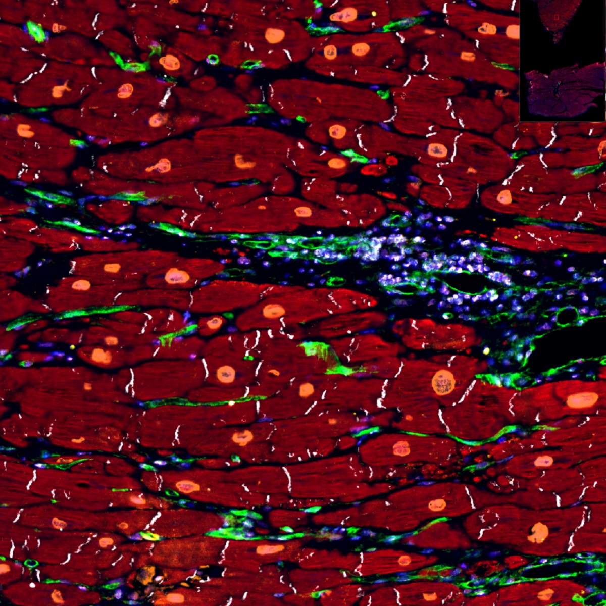
Understanding Hidden Inflammation in Heart Disease
Using cutting-edge techniques that allow us to map proteins and cells within heart tissue at high resolution, our research is uncovering how inflammation spreads and affects different regions of the heart—even in areas that appear normal under a microscope. These insights are helping us identify new markers of disease and may lead to better diagnostic tools and targeted therapies. Ultimately, this work aims to make invisible disease visible—and more treatable.
Collaborating for Cardiovascular Care